Categories
Colorectal General

Why is my child constipated?

This is the most common question in constipation clinic!

The answer for most families is IDIOPATHIC…which is code for “We can’t really explain this.” Unsatisfactory? You bet.

Instead, a simple framework sorts constipation into distinct patterns that can be understood. Remember, it’s J.U.S.T. constipation.

What does J.U.S.T. stand for?
  • J is for Juvenile: In the first year of life, babies undergo numerous changes and are still developing. Stool patterns can be wildly unpredictable. The key to this phase is supported bowel training, in the same we ‘sleep train’, support feeding, walking and language acquisition.
  • U is for Unexplained: Yes, for some children we never quite find a rational explanation. The strategy here is accepting the tendency towards constipation and managing well with diet, medications etc.
  • S is for Syndromic: There is an overarching diagnosis that comes with a tendency to constipation e.g.
    • Children with developmental delay on long term tube feeding have a restricted, low residue diet
    • Children on the autism spectrum may have sensory issues that hinder defaecation dynamics, or restricted diets that pre-dispose to constipation
  • T is for Transitional: Seen in toddlers and older at times of transition e.g. home to nursery or school, toilet training, travel. Withholding behaviour , prolapse and anal fissures are common in this group. The key for this group is breaking the cycle, establishing a bowel routine and long-term dietary hacks.

Categories
Colorectal General

Constipation: biopsy?

Constipation is a common problem in childhood.

Very rarely, an underlying and serious problem i.e. Hirschsprung’s disease, can be detected on biopsy.

Importantly, most biopsies done are actually negative.

However, it can be tricky moving forward with recommended treatments if there is an outstanding concern.

For parents, it can be quite difficult to decide if the time has come to discuss a biopsy with your paediatrician.

Paediatric surgeon’s are best placed to review and advise. We manage Hirschsprung’s disease and will have a good sense of whether a biopsy is needed or not.

Here are some facts about rectal biopsies for constipation.

Categories
Colorectal

Haemorrhoids

Yes, children can get haemorrhoids.

It’s rare, which is why reviewing the issue with a GP, then seeing a paediatric surgeon is advised.

Haemorrhoids / piles typically start of as a small lump that appears at the anus during stooling. When small, but visible (Grade II), they pop back in promptly after stooling.

As they get bigger (Grade III), they may stay out longer, and sometimes need a helpful hand popping back in. Your child may have some symptoms and signs at this stage e.g. bleeding / blood spotting from the haemorrhoid, itchiness, a feeling of sitting on a hard pea.

Large haemorrhoids are always out, and cannot be pushed back. They can get painful or clotted.

What causes haemorrhoids in children?

It’s not clear why children get haemorrhoids. However, we do tend to see them more in children and adolescents with constipation, who are spending time straining on the toilet. Once the constipation and straining , is resolved, there’s every chance the haemorrhoid improves without surgery.

Rarely, the haemorrhoid may be a sign of congestion arising from pressure from abdominal organs e.g. liver congestion, pelvic mass. Therefore, history and examination by a specialist is helpful. If in doubt, an ultrasound of the abdominal organs and domain can settle any outstanding worries, and allow confident watchful waiting.

What treatments are available?

For chronic, high-grade haemorrhoids , medical options are numbing creams and shrinking creams e.g. Anusol and Scheriproct.

Surgical options include:

  • Sclerotherapy
  • Haemorrhoidal artery ligation (HALO) procedure
  • Haemorrhoidectomy / excised haemorrhoidectomy
  • Sutured mucopexy

All these options involve looking inside the bottom with stretching instruments e.g. proctoscope, ultrasound probe etc. In children, due to their much smaller structures, there is a risk to damaging the anal spincter muscle. This is the muscle that helps us keep stools in and be continent. Therefore, you will find paediatric surgeons will want to try all non-operative options first, before offering surgery.

Sclerotherapy involves injecting the engorged blood vessels with an agent that causes inflammation and scarring. Various agents are used e.g. phenol, polidocanol, etc. This option can work, but may need more than one treatment, and may risk ulceration if the agent leaks into surrounding tissues.

HALO involves finding the feeding vessel and blocking it off with a stitch or clip. In adults, this can be done handily with ultrasound guidance. In children, a general anaesthetic would be needed and the child would need to be old / big enough to accept the ultrasound probe to the anus without injury. It can also be done under the surgeon’s direct vision, with careful stretch of the anus.

Sutured mucopexy is a similar procedure, where the artery feeding the haemorrhoid is pinched by a line of sutures running through the bowel wall.

Typical recurrence rates are 10-20% for these surgical procedures.

Excision haemorrhoidectomy removes the haemorrhoid all together, and recurrence after this procedure is less common. However, the child needs to be big enough to accept the stretching instruments required to get a really good view of the haemorrhoid.

Laser treatment is a good option for adults, but we don’t have enough experience in children to be make recommendations as yet. Its best to ask your surgeon about their experience with this method.

Consulting a paediatric surgeon and finding out which procedure they would recommend for your child and setting is key.

Categories
Colorectal

Sacral Agenesis

Type I-IV

I : Unilateral, partial or total

II: Bilateral, partial, symmetrical

III: Total sacral agenesis, iliac bones articulated lateral to last lumbar vertebra. Variable lumbar agenesis

IV: Complete sacral agenesis with iliac bones fused beneath last lumbar vertebra
Categories
Colorectal General

Constipation: when do you need a biopsy?

Constipation is a common problem in childhood. Very rarely, an underlying and serious problem i.e. Hirschsprung’s disease, can be detected on biopsy. Importantly, most biopsies done are actually negative.

For parents, it can be quite difficult to decide if the time has come to discuss a biopsy with your paediatrician.

Here are some facts about rectal biopsies for constipation.

Categories
Colorectal

Rectal biopsy for constipation: a guide for parents

Constipation is a common problem in childhood. Most children will have an episode that passes naturally, or with changes in diet and perhaps some medication. However, some children will have chronic, ongoing issues that escalate, needing surgical intervention.

When is a biopsy of the rectum useful?

If 10 children have a biopsy, 1 or 2 may have a positive finding of Hirschsprung’s disease.

However, a review will be necessary before the biopsy to find out if there are other risk factors.

NICE recommends that a rectal biopsy should not be performed unless there is:

-delayed meconium (>48hrs after birth in a term baby

-early constipation (first weeks of life)

-chronic abdominal distension plus vomiting

-family history of Hirschsprung’s disease

-faltering growth, in addition to any of the previous features

Other features have been suggested:

vomiting, in addition to abdominal distension is a risk factor for Hirschsprungs

-a rectal biopsy is more likely to be negative with increasing age

girls are less likely to have a positive biopsy for Hirschsprung’s

How is the biopsy done?

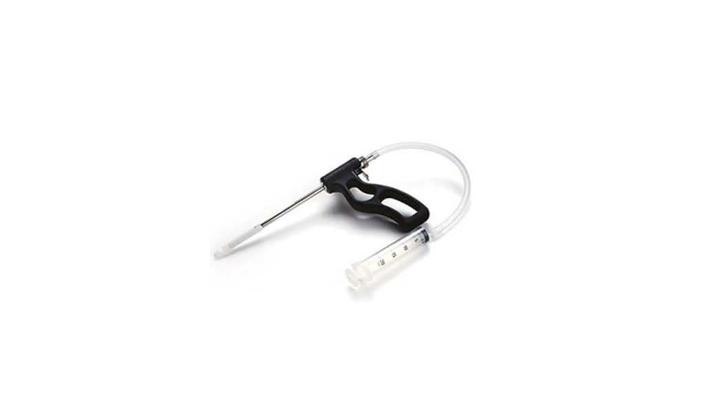
Suction biopsy: A small , finger-like biopsy tube is placed inside the rectum. A syringe is used to suck some of the rectal tissue into the tube, which has a cutting mechanism. A small sample is pinched off. As the sample is small, the surgeon may choose to take a 2-3 samples at different levels, to increase the chance of a clear result. Most doctors would like to do this under the age of 6 months.

This method can be carried out in an awake baby, as long as can be effectively wrapped to keep them still. Over 6 months, the baby is strong enough to move, making the test more difficult to achieve effectively.

Strip biopsy: This method involves a general anaesthetic. Once asleep, the surgeon looks inside the rectum and cuts out a 1-2cm strip of tissue. 

Regardless of method, these samples are then sent to the lab. 

What are the lab looking for?

In summary, nerve fibres and ganglion cells.

Ganglion cells are specialised hubs where nerve fibres meet and communicate. The histopathologist in the lab looks to confirm the presence of the nerve fibres and ganglion cells.

In Hirschsprung’s disease, there are no ganglion cells (hence the term- aganglionosis) within the bowel layers.

Nerve fibres are absent. Abnormally thickened nerve trunks are also observed.

This ganglion cell test is very sensitive for Hirschsprung’s disease. If the ganglion cells are seen, your surgeon can be >93% sure that your child doe not have Hirschsprung’s disease.

Categories
Colorectal

Hirschsprung’s disease

An uncommon but important cause of neonatal bowel obstruction.

Definition

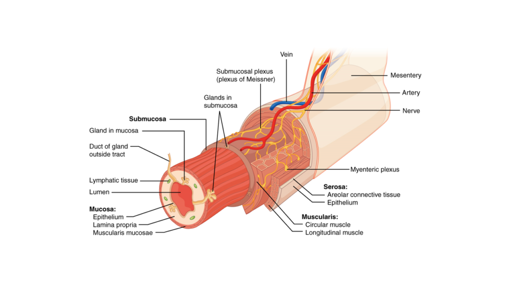
Hirschsprung’s disease (HSCR) is an aganglionisis of the distal gut due to failure of migration of neural crest cells to myenteric and submucosal bowel pexus 

Presentation

HSCR can present differently depending on age and severity.

Antenatal suspicion

Neonatal period

  • constipation progressing to obstipation
  • abdominal distension
  • bilious vomiting
  • delayed passage of meconium

Hirschsprung’s associated enterocolotis (HAEC)

  • constipation with spurious/ overflow diarrhoea
  • abdominal distension, with visible , palpable bowel loops
  • failure to thrive
  • pyrexia

Age at diagnosis varies, and is likely dependent on robustness of health systems and neonatal screening programmes.

  • In US, >70% neonatal diagnosis
  • In NW Tanzania: ~5% neonatal presentation
Age and Incidence

Rare, 1 in 3500 live births .

Antenatal

Neonatal

Childhood

Adulthood

Sex

Male > Female

Genetics

Most cases of HD are sporadic (80%)

Where there is a pattern of inheritance (20%), the association can be syndromic or familial

Syndromic HD

  • Trisomy 21 :
    • 3 percent of T21 patients have HSCR.
    • 7% of HSCR patients have T21.
  • Neurocristopathies (disorders of neural crest cell differentiation and migration)
    • Shah Waardenburg syndrome type IV
    • Mowat-Wilson syndrome: Microcephal, characteristic facies, GDD.  50% of MWS patients will have HD. Associated with ZEB2 gene mutations.
    • Congenital central hypoventilation syndrome (Ondine’s curse) : autonomic dysfunction syndrome predisposing to sleep apnoea, HSCR (~15%, Haddad syndrome) and neural crest tumours (5-10%). Associated with PHOX2B gene mutations.
    • Heriditary neuuroblastoma with Hirschsprung’s disease syndrome (PHOX2B  mutation)
    • Multiple endocrine neoplasia : MEN2B (RET, GFRA genes). Ganglioneuromas are included in the spectrum of intestinal neuronal dysplasia (IND) . Association with medullary thyroid carcinoma ( RET-620 (the “Janus gene”).
    • Heriditary neuuroblastoma with Hirschsprung’s disease  (PHOX2B related)

Familial HSCR

  •  EDNRB and RET gene mutations implicated
  • Progressive penetrance with implications for aganglionic length i.e. increased risk of long segment, total colonic and total intestinal presentations
  • Autosomal pattern of inheritance, but penetrance is variable
  • Parental transmission ~20%, Sibling risk ~ 10%


Aetiology

Differentials

For neonates presenting with infrequent of delayed passage of stools, consider

  • anorectal malformation
  • small left sided colon syndrome
  • meconium plug syndrome
  • meconium ileus

Variants of HSCR: These are conditions with a similar presentation to HSCR, but with ganglion cells identified on rectal biopsy.

Variants described include:

  • megacystis microcolon intestinal hypoperistalsis syndrome
  • intestinal neuronal dysplasia
  • intestinal ganglioneuromatosis
  • isolated hypoganglionosis
  • immature ganglia
  • absence of the argyrophil plexus
  • internal anal sphincter achalasia

Diagnosis should be based on histo-immunochemistry and electron microscopy.

Severity/ Staging

HSCR is staged in a segmental approach.

Ultrashort: within 3cm of anus i.e. distal rectum. Characterised by late presentation in childhood.

Short segment/ Rectosigmoid: This is the most common i.e. 75%

Long segment: Involving colon proximal to the sigmoid. ~20%

Total colonic aganglionosis (TCA): ~4%

Total Intestinal ganglionosis (TIA). ~1%. This cohort is unusual due to:

  • early presentation with delayed passage of meconium (d4-8)
  • collapsed bowel, little or no intestinal distension, no transition zone
  • hypertrophic nerve fibres may be absent
  • seen more commonly in familial HSCR

Investigations

Bedside

  • Abdominal examination
  • Digital rectal examination / anal calibration

Blood tests

  • CF testing for delayed passage of meconium
  • Neonatal thyroid function screen

Specimen

  • Rectal biopsy :
    • In neonates and younger infants , this can be achieved by the bedside using blind rectal suction. Biopsy forceps include rbi2, Noblett, solo-RBT, Scheye.
    • In older infants and children, a general anaesthetic is indicated under direct or endoscopic vision. Strip or cup biopsies are taken.
    • Histopathology
      • Dual approach of histopathology (H&E) and immunohistochemistry (Calretinin)
      • H&E is 85% specific and 100% sensitive
      • Calretinin is 97% specific and 100% sensitive

Imaging:

Contrast enema can be contributory towards an HSCR diagnosis.

Special tests:

  • RAIR:
    • Recto-anal inhibitory reflex is tested on anorectal manometry
    • With a false negative rate of 4%, it is contributory toward ruling our HSCR. However, the gold-standard diagnostic i.e. Calretinin immunohistochemistry has 100% sensitivity and 97% specificity. Considering the consequence of a missed HSCR diagnosis, RAIR may be considered contributory but non-diagnostic.

Controversies

Primary versus staged approach

Age at biopsy


Categories
Colorectal

Anal Fissure

Sorry, but only verified contributors, editors and curators can view this content.

If you are a health professional, please sign up and get verified to view this content. Members are verified within 24 hours of sign-up.

Parents and caregivers: Advanced content contains deep-dive information which sometimes needs protection for propriety or professional reasons. Please respect this restriction. We will always try to provide as much high-quality information as possible, within these bounds. If you would like us to make a page more accessible , please email the link to editor@psionjournal.org for review.


Categories
Colorectal

Bloody stool

In toddlers and little children, anal fissures are the most common reason for blood on stools.

An anal fissure is a superficial tear in the anus skin. It results in painful stooling, and blood on the stools, on wiping or in the nappy.

How does an anal fissure develop?

These tears arise often after an episode of constipation. Passing a large hard stool initially stretches the anus skin margin. This stretches the skin creating a little tear. As subsequent hard or large stools pass, they are painful to do. Your child may then, understandably, start withholding. This involves behaviours that try to avoid that the pain associated with passing stools. This worsens the constipation and sets up a vicious cycle of stooling and tearing.

What does an anal fissure look like?

The tears typically appear along the midline. Imagine the anus is a clock-face.

Lying down, the genitals mark 12 o’clock and the back mark’s 6 o’clock. These tears appear at 12 and 6 o’clock.

The tears themselves can be hard to see. However, there is often a little lump below them, where the skin has thickened from trying to heal over repeatedly. This little lump is called a skin tag. It is a sentinel marker , or sign, that there is, or was, an anal fissure in that area.

If there are multiple tears in a sun-ray pattern, this is unusual, and needs review by a doctor.

What can be done to treat anal fissures?

It can take several weeks to treat an anal fissure. The key thing to do is to break the cycle of hard/ large stools and tearing. So, although your baby or child may not seem to be constipated (passing infrequent stools), it is important to ensure they are passing soft stools for a few weeks. This allows the fissure area to heal.

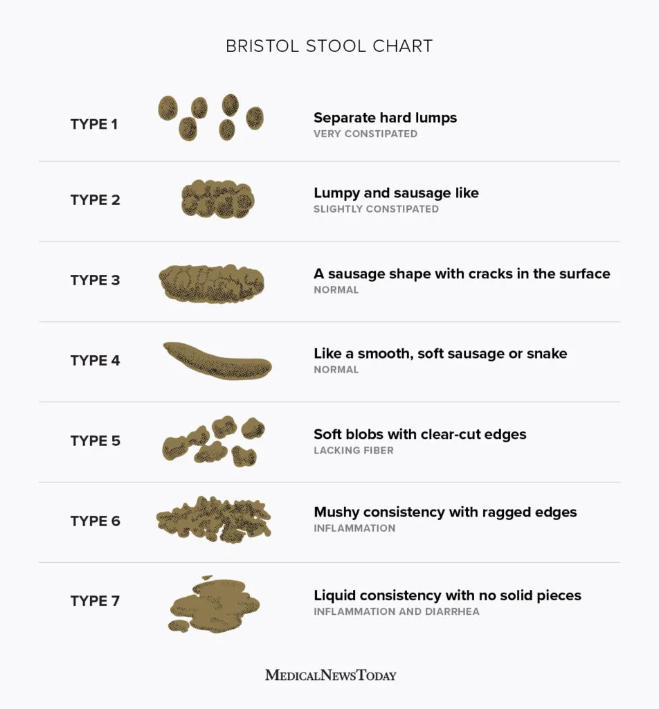
The Bristol stool chart is a useful tool for rating stool consistency.

Bristol Stool Chart

When your child has an anal fissure, aim for stools that are type 4 and above.

This should be done primarily with adequate fluid intake and a diet high in fibre. If further softening is required, stool softening laxatives can be considered. For more information, read about Diet and Constipation.

What about applying a cream?

There are different creams that your doctor will consider.

Soothing creams e.g. scheriproct contain some local anaesthetic and some steroid. It may not be suitable for all babies and children and must be prescribed.

Vasodilator creams work by increasing blood supply to the area. This include diltiazem and GTN. However, these are only prescribed after careful consideration with your surgeon. There may be side effects to think about, particularly if treatment requires several weeks.

Are there surgeries for anal fissure?

Yes. Your surgeon may recommend botox therapy to the anal sphincter. This treatment also has side effects to consider e.g. incontinence, recurrence.

You may have read about other, more radical surgical treatments e.g. fissurectomy, spincterotomy. These treatments are described for adult patients.

Other causes of bloody stools

Blood on or mixed in with stools can also be due to:

  • Allergies e.g. cow’s milk protein allergy in babies
  • Bacterial diarrhoea
  • Intussusception
  • Bleeding Meckel’s diverticulum
  • Inflammatory bowel disease e.g. ulcerative colitis or Crohn’s
  • Polyps

Your GP is best placed to review and refer to an appropriate specialist, if needed.

Categories
Colorectal

Bowel problems

The paediatric colorectal surgeon deals with an varied and challenging mix of conditions. These conditions present at different ages, creating an interesting case-mix of neonatal, paediatric and adolescent patients.

Neonatal presentation

  • Anorectal malformation
  • Hirschsprung’s disease

Childhood presentation

  • Constipation and feacal incontinence
  • Anal fissure

Adolescent presentation

  • Inflammatory bowel disease
  • Pilonidal disease
Creative Commons License
Except where otherwise noted, PSIONjournal by Open Knowledge Platforms is licensed under a Creative Commons Attribution-NoDerivatives 4.0 International License.